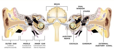
Anatomy & Physiology of the Hearing System
The human auditory system consists of four parts, which are:
1) External ear
2) Middle ear
3) Inner ear
4) Central nervous pathways
Anatomy and Physiology of the external ear
The external ear consists of two parts: earlobe, external auditory canal 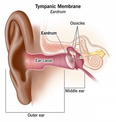
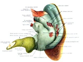
Auricle
The earlobe is made of elastic cartilage, which is covered by skin, and the earlobe cartilage extends to the outer third of the external ear canal, but the eardrum does not have cartilage.
Ear lobe muscles
There are muscles inside the earlobe whose mobility is not noticeable in humans. The number of these muscles is 3, which are the upper, anterior and posterior muscles that connect the earlobe to the skull.
Earlobe function
Gathering of voices
Orientation and localization of sound due to collision with asymmetric components of the earlobe
Resonance and amplification of sounds, especially at high frequencies
Localization
The shape of the edges and ridges of the earlobe causes amplification and resonance of the sound in high frequencies (due to the short wavelength of these frequencies) around 5 kHz, the resonance pattern of these frequencies changes according to the location of the sound source, so the earlobe is in the direction Orientation is effective on the horizontal level. It seems that the earlobe is also effective in orientation on the vertical level (in humans).
External auditory canal
On average, the length of the ear canal of adults is 2.5 cm and its diameter is 0.7 cm. The channel is slightly oval. The outer one-third to one-half of the canal has a flexible cartilage frame, which is very similar to the rest of the external ear. The remaining part, i.e. the inner two-thirds of the internal external auditory canal, is connected to the temporal bone of the skull. The diameter of the canal decreases from the outside to the inside, and increases to a minimum in an area called the isthmus. The external ear canal lies in an S shape and the presence of this curvature in the canal makes it difficult to see the tympanic membrane.
There are two types of cells in the cartilaginous part of the canal to secrete cerumen.
Sebaceous glands
Cerumen glands
These glands perform protective and antibacterial action. Cerumen is transferred to the outside by the movement of the skin of the duct
And there is no need to use ear cleaner. Cerumen secretion increases with age, especially in men. 45 decibel hearing loss occurs with compressed cerumen.
The skin of the ear canal migrates outward and clears the ear canal and tympanic membrane of dried cerumen and other small waste particles. In addition to cleaning the canal from small objects, the migration of the canal skin is also involved in the spontaneous and rapid healing of tympanic membrane tears.
Physiology of external auditory canal
The outer ear is a band-pass filter that emphasizes high frequencies. By creating an obstacle in the external ear opening, it becomes a low-pass filter. The ear canal, like closed vocal tubes, amplifies sounds by 10 to 15 decibels in the frequency range of 3 to 4 kHz. In addition, the external ear canal along with the earlobe play a role in the direction of sounds.
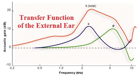
The reinforcing effect of the tube and the external ear canal and the eardrum in total exceeds 20 decibels. It seems that in the frequency range of 1.5-7 kHz and 2-5 kHz, the tube has an amplification effect more than 5-15 dB and 20 dB, respectively.
Anatomy and function of the middle ear
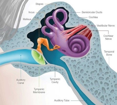 The middle ear is a cavity filled with air and covered with mucus between the tympanic membrane and the inner ear and includes the tympanic membrane, three ossicles Hammer, anvil and stapes, ligaments and muscles related to bones and Eustachian tubes. The tympanic membrane is a semi-transparent, oval-shaped membrane at the end of the external auditory canal. The weight of the curtain is 14 grams and its long diameter is 9-10 mm and its short diameter is 8-9 mm. The tympanic membrane is placed in the shape of a cone at the beginning of the bony chain, so that the handle of the hammer bone is placed on top of the membrane, and this condition makes the membrane and the chain work together, and at the same time, this integration causes the sound pressure to increase. In addition, it should be kept in mind that the effective surface of the eardrum is 17 times the surface of the stapes bone, which is located at the end of the bone chain and on the oval valve.
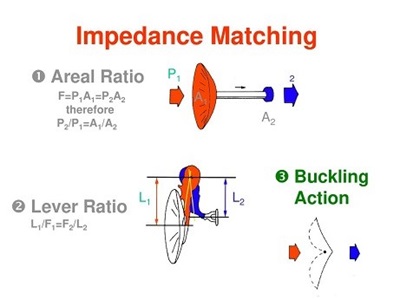
In addition, the ossicles of the middle ear increase the sound pressure in the
bottom of the last ossicle (stipe) through a lever function, and in this way the energy loss/sound pressure loss caused by the transfer of sound from air to liquid is compensated. Therefore, in fact, the function of the middle ear is to match the air resistance of the outer ear and the resistance of the inner ear fluid. In fact, the function of the middle ear causes sound energy to enter the inner ear fluid without loss from the outer ear space.
Anatomy and physiology of the inner ear
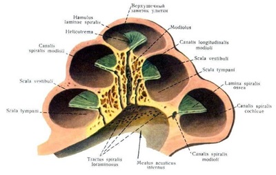
The cochlea is necessary to hear sounds. The cochlea has sensory cells that are responsible for converting mechanical waves into electrical activity that is transmitted to the brain and perceived as sound.
The damage of these sensory cells can be seen due to loud noise, some drugs, inner ear infection or aging. Their permanent loss is characterized by sensorineural hearing loss. The middle and inner ear are completely inside the very hard part of the temporal bone, which is called the spine. In fact, the bony labyrinth is a part of the tympanic bone that covers the inner ear.
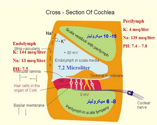
The bony labyrinth was created to protect and support the end organs of hearing and balance. Inside the bony labyrinth, there are membrane parts that include cells and fluids of the inner ear, which are responsible for converting the mechanical energy of sound into electrical energy. The inner ear is connected to the middle ear through round and oval valves.
In the inner ear, after converting the mechanical current into an electrical current, the ends of the afferent nerve fibers are stimulated, and then the electrical current is directed to the brain through the auditory nerve fibers in the central nervous system, and the sound is heard.
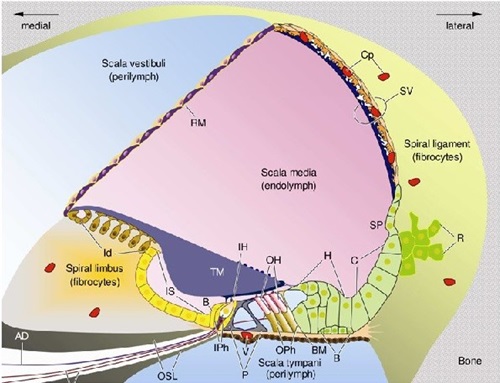
Physiology of the Inner Ear
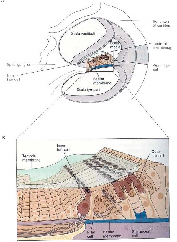
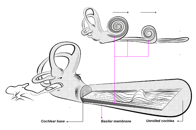
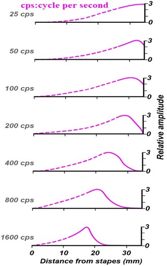
By entering mechanical energy into the cochlea and transferring energy in the cochlea in the form of a traveling wave and receiving this mass of energy in a certain area of the cochlea based on the matching of the frequency of the carrier wave with the characteristics of the basilar membrane (the length of the transverse fibers and their hardness), it causes the maximum displacement in the basilar membrane. The division of the basement membrane in the cochlea is called tonotopic organization, which was first comprehensively proposed by Von Bekesy.
The movement of the basilar membrane causes the cilia of the external hair cells to bend, which in turn will stimulate the external hair cells in the same area, and the stimulation of the external hair cells will result in the shortening of the size of these cells.
The tip of the cilia of the external hair cells is pulled down, and as a result, the tip of the cilia of the internal hair cells is bent, and this bending causes the opening of the ion valves at the tip of the cilia of the inner hair cells, and the cell is stimulated.
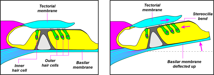
The stimulation of the inner hair cells causes the release of neurotransmitters at the base of the inner hair cells, and as a result, the afferent fibers of the auditory nerve are stimulated, and the
electric current in the nerve fibers is transferred to higher centers in the auditory nervous system and finally reaches the brain, and hearing occurs in the brain.
Central Auditory Nervous System (CANS)
After converting the sound energy into electric current in the inner ear, the fibers of the auditory nerve (cranial nerve) send the electric current in coded form to the centers and stations in the central nervous system. In addition to transmitting audio information, the auditory pathways provide information about the power and position of the sound in space to the presenting brain. In fact, our ear transmits the sound stimuli entered through the outer ear to the middle ear and then to the inner ear, and in the inner ear, the mechanical waves are converted into electrical waves. The stimulation of the nerve fibers creates neural codes that the brain can decode, process and understand, and as a result sounds are heard.
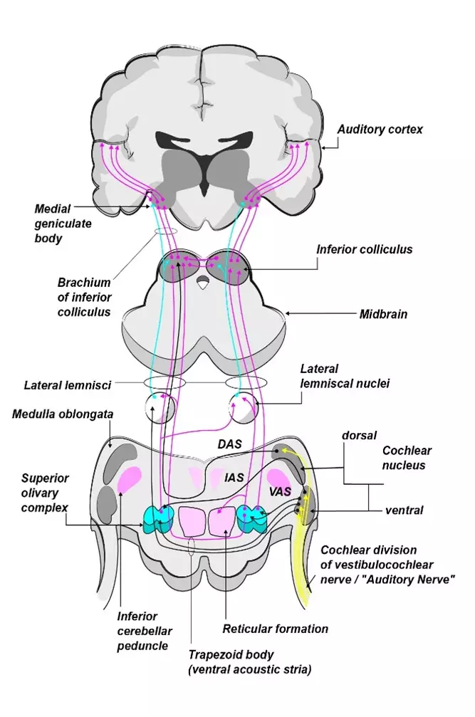
Information from the auditory nerve is then sent to the brain via several nuclei in the brainstem (cochlear nuclei, superior olivary nucleus, lateral lemniscus, inferior colliculus) and midbrain (medial geniculate body). These cores allow further processing of the audio message.
In a quick look at the auditory central nervous system, the following sections can be mentioned as the main stations on this path, which are:
1- The cochlear nuclei
are the first auditory information relay center in the brain stem. The cochlear nucleus includes the dorsal cochlear nucleus and the ventral cochlear nucleus, both of which receive input from the auditory nerve, but the dorsal cochlear nucleus is higher. The ventral part of the cochlear nuclei is divided into two anterior and posterior parts, thus creating three parts in the cochlear nuclei, and in the same way, three groups of ventral, medial and dorsal nerve fibers leave the cochlear nuclei, which send auditory information to higher centers. The distal and proximal parts of the auditory nerve along with the cochlear nuclei are the first centers in the auditory pathway that produce measurable electrical signals from the scalp.
2- The Superior Olivary Complex
that plays special roles in the hearing of two ears. The superior olive complex includes the medial and lateral superior olive nuclei. These nuclei are hubs of neurons that receive input fibers from the dorsal and ventral cochlear nuclei and send output fibers to the nuclei of the lateral lemniscus. The superior olivary nuclei are the first nuclei to receive input from both ears, making the superior olivary complex important for binaural hearing. The medial olivary nuclei (MSO) contain more neural representation of low frequencies while the lateral olivary nuclei (LSO) have more connections for high frequencies. MSO compares low-frequency wavelengths to calculate sound arrival time differences between the two ears (interaural time differences ITDs), while LSO compares high-frequency wavelengths to calculate sound level differences between the two ears (Differences in sound intensity between two ears ILDs).
3- Lateral lemniscus
where major information exchange between two auditory pathways takes place in this station. The lemniscus nuclei have three parts: ventral, dorsal, and medial, and although the function of this station is not precisely known, it seems that this station is sensitive to amplitude modulation.
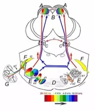
4- The inferior colliculus complex
is one of the most important auditory information relay centers in the auditory system, which is located in the midbrain, and in some sources, its location is in the highest part of the brain stem. The roles of inferior colliculus include mutual communication between two auditory paths (right and left), orienting to new stimuli, providing feedback to auditory efferent fibers, and detecting and tracking amplitude and frequency modulation. Also, the reception of tactile information and the initial integration of visual and auditory information takes place in this station.
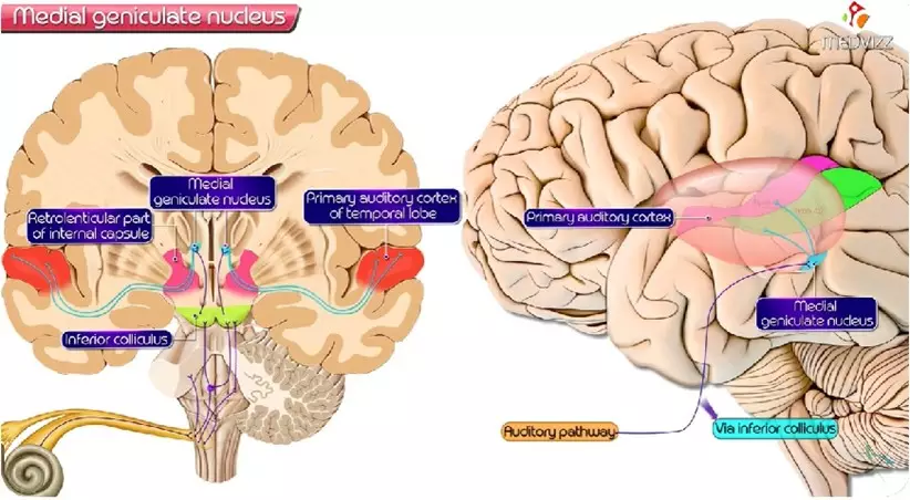
5- Medial Geniculate Body
located in the midbrain and in the thalamus and is the last relay center for auditory information before reaching the auditory cortex. The thalamus collects sensory inputs from various modalities and transmits them to the cerebral cortex. The medial knee body includes a segment that has a tonotopic organization. The main function of the internal geniculate body is to transmit auditory information to the cerebral cortex in addition to integrating sensory information.
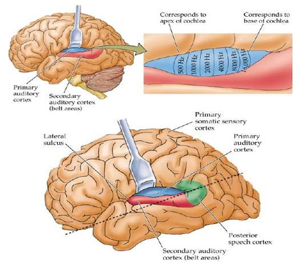
6- Primary auditory cortex
The auditory cortex includes the primary and secondary auditory cortex. The primary auditory cortex is also known as A1 and is located in the temporal plane, marked by the Sylvian fissure on the upper surface of the temporal lobe. The secondary auditory cortex (A2) surrounds the primary auditory cortex.
The primary auditory cortex has a tonotopic organization such that the lateral edge of the Sylvian fissure is sensitive to lower frequencies and the inner part of the cortex is sensitive to high frequencies. The primary auditory cortex is responsible for perceiving pitch, rhythm, and duration of sound, while the secondary auditory cortex is responsible for pattern perception, which includes perceiving speech and melody.
Anatomy and physiology of the vestibular system
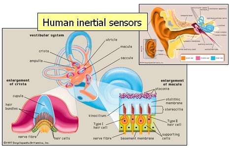
Recognition of movement and sense of position in space are carried out by vestibular receptors. Vestibular receptors convert the information related to the direction and speed of movement as well as the position of the body into electrical current and send it to the brain. The information sent to the brain is combined with visual, positional and movement information from the cerebellum and cerebral cortex, and as a result, controls gaze, body posture, automatic reflexes, and spatial orientation, which is mostly done on a non-conscious level. become It should be noted that movement is perceived, but there is no primary vestibular cortex, and the part of the cortex that does this work also receives information from other senses, including vision and visual perception. In fact, in daily life, the vestibular system, in addition to constantly understanding and identifying the position of our body in space in relation to gravity, also recognizes rotational and linear movements and provides appropriate responses to them.
Therefore, in the vestibular system, there are receptors for the sense of gravity and other linear accelerations, as well as receptors for angular accelerations, and in addition, it regulates the tonicity of the body muscles by sending nerve messages. The vestibular system works based on the balance between the left and right ears.
The vestibular nerve continuously sends signals to the brain that this rate of sending signals while resting (resting potential) causes the recognition of the state of the body, and during the movement and activity of the body, the increase of nerve signals to the brain and their processing leads to the understanding of the new state. and the brain issues the necessary commands to the muscles and organs of the body according to the received signals and combining them with the information of other senses. The vestibular system has 5 sensory receptors, which are: 3 sensory receptors in the horizontal, vertical and posterior semicircular canals that respond to rotational movements in 3 spatial axes.
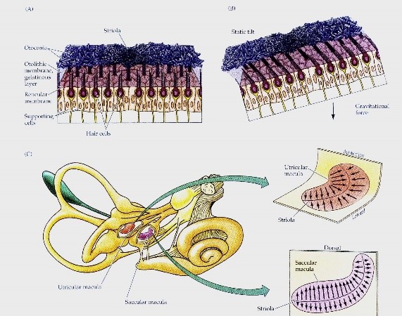
Also, 2 otolith organs in saccule and utricle which respond to linear movements in vertical and horizontal directions. The vertical canals and saccule are located in the vertical axis and the horizontal canal and utricle are located in the horizontal axis of the head. These organs establish neural connections in the brain with the oculomotor system (movement of the eyes) and position control. In addition to sensory receptors, there is also fluid in the vestibular system, which moves the fluid to transfer pressure and cause stimulation in the sensory receptors. As a result, mechanical energy is converted into electrical energy which stimulates the nerve fibers and finally receives electrical information in the brain and processes and integrates them with information from other senses to establish balance.